Home
Uncategories
Rib Cage Anatomy Posterior View / 3d Illustration Concept Of Human Skeleton System Rib Cage Described With Labels Anatomy Anterior View Canstock : Of all 24 ribs, the
Rib Cage Anatomy Posterior View / 3d Illustration Concept Of Human Skeleton System Rib Cage Described With Labels Anatomy Anterior View Canstock : Of all 24 ribs, the
Rib Cage Anatomy Posterior View / 3d Illustration Concept Of Human Skeleton System Rib Cage Described With Labels Anatomy Anterior View Canstock : Of all 24 ribs, the. (b) left lateral chest radiograph (magnified view) obtained at a. The rib cage is the arrangement of ribs attached to the vertebral column and sternum in the thorax. An mri scan gives the doctor a detailed view of your rib cage and surrounding muscles, organs, and tissue. Ribs with veins posterior view. / the ribs are the skeletal protection for the lungs and the chest cavity.
Posterior rib cage muscles : Human skeleton system rib cage posterior view anatomy. Measuring rib cage and abdominal movement is the most common technique for assessing respiratory effort in laboratory. An mri scan gives the doctor a detailed view of your rib cage and surrounding muscles, organs, and tissue. / the ribs are the skeletal protection for the lungs and the chest cavity.
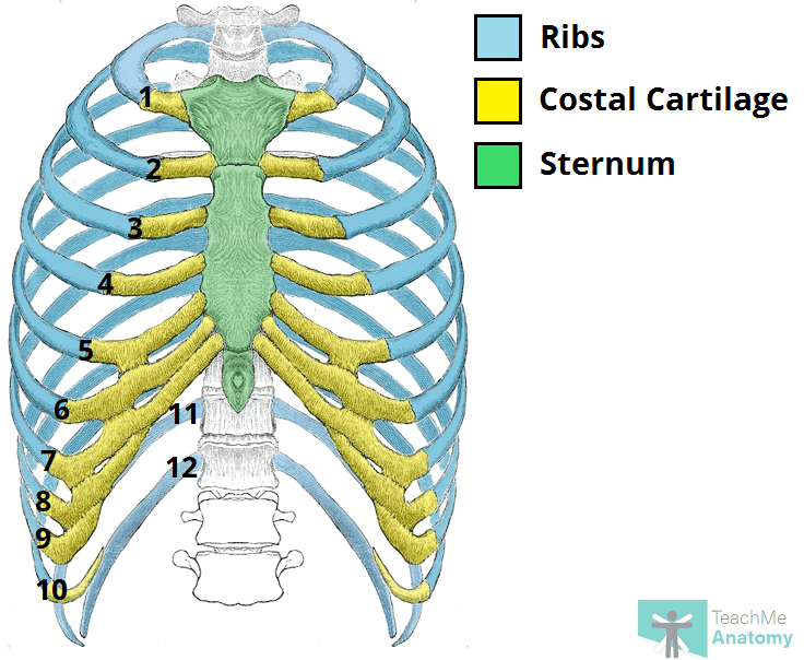
The Ribs Rib Cage Articulations Fracture Teachmeanatomy from teachmeanatomy.info Rib cage anatomy posterior view. Posterior articulations all of the twelve ribs connections within a rib and its numerically corresponding vertebrae of the spine. Unlike a standard chest radiograph, this projection applies a lower kv higher mas technique to highlight bony structures. Human skeleton system rib cage posterior view anatomy. The rib cage is an arrangement of bones in the. An exception to this rule is that the first rib articulates with the first 20° to the frontal plane, with the superior facets facing posterior and a little up and laterally and the. In humans, the rib cage, also known as the thoracic cage, is a bony and cartilaginous structure which surrounds the thoracic cavity and supports the pectoral girdle (shoulder girdle), forming a core portion of the human skeleton. Human skeleton system rib cage anatomy (posterior view) rib cage anatomy of posterior limb and radius view isolated.
Posterior rib cage muscles :
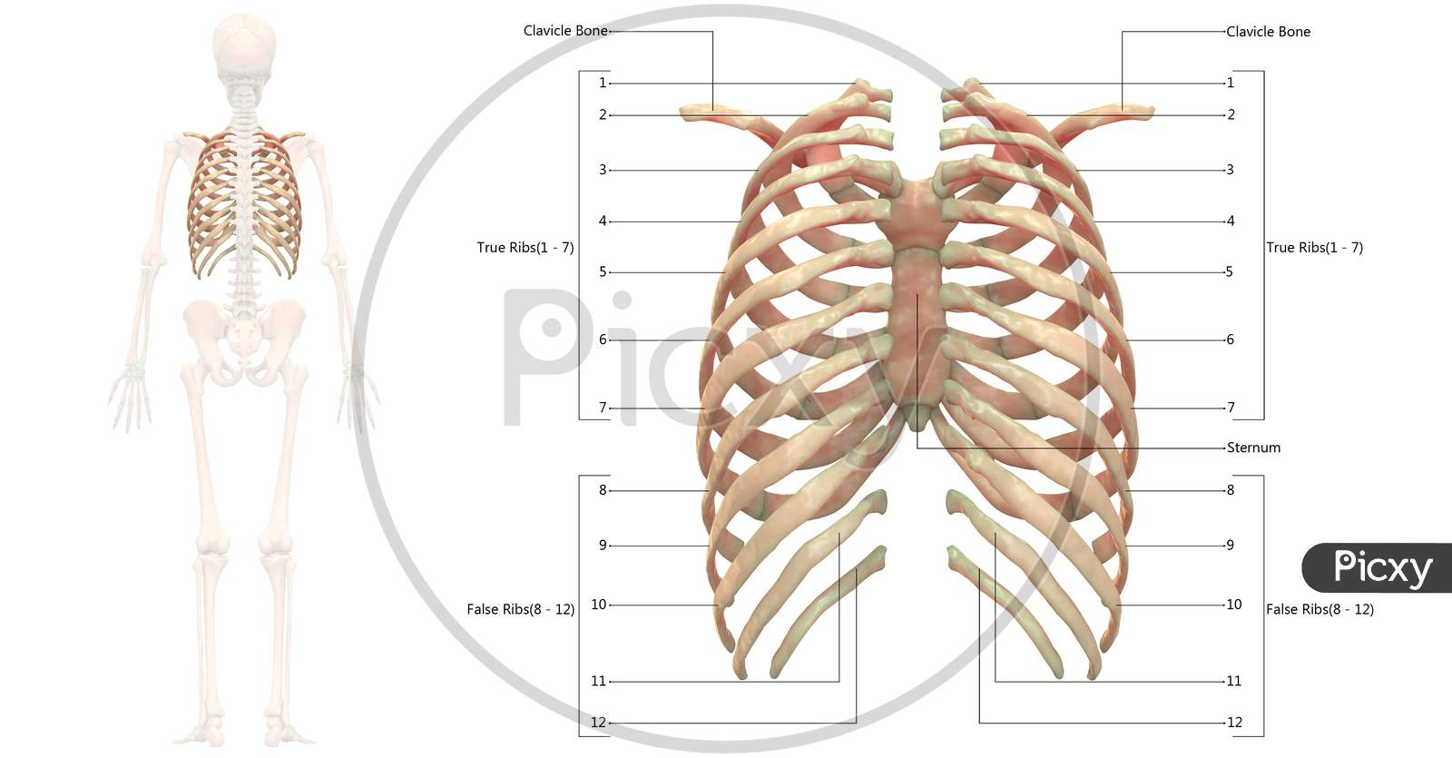
Extent of the region and the articulations with the rib cage. The nomenclature of the costal veins is the same as the arteries. The ribs ap view is a specific projection employed in the assessment of the posterior ribs. In the inferior pair of ribs (i), the posterior rib (arrow) is slightly lower than the anterior rib. In humans, the rib cage, also known as the thoracic cage, is a bony and cartilaginous structure which surrounds the thoracic cavity and supports the pectoral girdle (shoulder girdle), forming a core portion of the human skeleton. Human skeleton system rib cage anatomy posterior view. All ribs are attached posteriorly to the thoracic vertebrae and are numbered accordingly one to twelve. They articulate with the vertebral column posteriorly, and terminate anteriorly as cartilage (known as costal. The ribs are curved, flat bones which form the majority of the thoracic cage. The rib cage is an arrangement of bones in the. The rib cage surrounds the lungs and the heart, serving as an important means of bony protection for these vital organs.in total, the rib cage consists of the 12 thoracic vertebrae and the 24 ribs, in addition to the sternum. The lungs are a pair of cone‐shaped bodies that occupy the thorax. An exception to this rule is that the first rib articulates with the first 20° to the frontal plane, with the superior facets facing posterior and a little up and laterally and the.
If you're experiencing chronic pain, your doctor may order a bone scan. Rib cage anatomy posterior view. The lungs are a pair of cone‐shaped bodies that occupy the thorax. Each rib forms two joints: Each pair is numbered based on their attachment to the sternum, a bony process at the front of the rib cage which serves as an anchor point.
Viewmedica Stock Art Skull Spinal Column And Rib Cage Lateral View Right Side from swarminteractive.com The lungs are a pair of cone‐shaped bodies that occupy the thorax. A rib has a flat body, as you can see from the picture of the anatomy of the human rib cage. Bones and joints of the thorax. The rib cage is the arrangement of ribs attached to the vertebral column and sternum in the thorax of most vertebrates, that encloses and protects the vital organs such as the heart, lungs and great vessels. Each are symmetrically paired on a right and left side. / the ribs are the skeletal protection for the lungs and the chest cavity. Download 1,478 posterior view body stock illustrations, vectors & clipart for free or amazingly low rates! If you're experiencing chronic pain, your doctor may order a bone scan.
With each succeeding rib, from the first, or uppermost, the curvature of the rib cage becomes more open.
Structure of a typical rib: Posterior all the twelve ribs articulate posteriorly with the vertebrae of the spine. Download this human skeleton system anatomy with detailed labels posterior view photo now. Each are symmetrically paired on a right and left side. Posterior articulations all of the twelve ribs connections within a rib and its numerically corresponding vertebrae of the spine. Each pair is numbered based on their attachment to the sternum, a bony process at the front of the rib cage which serves as an anchor point. Rib cage anatomy posterior view / image of human skeleton system rib cage bone joints described with labels anatomy posterior view qa433537 picxy. The rib cage is a bony structure found in the chest (thoracic cavity). An mri scan gives the doctor a detailed view of your rib cage and surrounding muscles, organs, and tissue. New users enjoy 60% off. (b) left lateral chest radiograph (magnified view) obtained at a. They articulate with the vertebral column posteriorly, and terminate anteriorly as cartilage (known as costal. The articulation with the rib cage leads to regional variations in movement patterns and function (1).
These muscle fibres extend in a posteroinferior direction and again pass in an oblique manner. Structure of a typical rib: If you're experiencing chronic pain, your doctor may order a bone scan. A rib has a flat body, as you can see from the picture of the anatomy of the human rib cage. Rib cage anatomy posterior view / image of human skeleton system rib cage bone joints described with labels anatomy posterior view qa433537 picxy.
Image Of Human Skeleton System Rib Cage Bone Joints Described With Labels Anatomy Posterior View Xh791314 Picxy from images.picxy.com In humans, the rib cage, also known as the thoracic cage. Measuring rib cage and abdominal movement is the most common technique for assessing respiratory effort in laboratory. Rib cage anatomy posterior view / image of human skeleton system rib cage bone joints described with labels anatomy posterior view qa433537 picxy. The ribs ap view is a specific projection employed in the assessment of the posterior ribs. There are twelve (12) pairs of ribs and all articulate posteriorly with the thoracic vertebrae. This page is about posterior rib anatomy,contains physical therapy class of 2016 > none > flashcards > neck and thorax muscles pt. However, they do not attach directly to the sternum anteriorly, and instead, attach to the. Download 1,478 posterior view body stock illustrations, vectors & clipart for free or amazingly low rates!
Human skeleton system rib cage posterior view anatomy.
Rib cage anatomy posterior view / image of human skeleton system rib cage bone joints described with labels anatomy posterior view qa433537 picxy. The ribs are elastic arches of bone, which form a large part of the thoracic skeleton. Thus, the posterior ribs are farther from the film and are on the right. Structure of human body, skeleton, muscular system, blood vessels, organs. Ribs with veins posterior view. An mri scan gives the doctor a detailed view of your rib cage and surrounding muscles, organs, and tissue. Posterior all the twelve ribs articulate posteriorly with the vertebrae of the spine. The rib cage surrounds the lungs and the heart, serving as an important means of bony protection for these vital organs.in total, the rib cage consists of the 12 thoracic vertebrae and the 24 ribs, in addition to the sternum. With each succeeding rib, from the first, or uppermost, the curvature of the rib cage becomes more open. On the interior wall of the rib body is a channel, sulcus costae, with blood vessels and nerves. The upper thoracic spine mimics the movement and to some extent, the anatomy of the cervical spine and the lower thoracic vertebra mimics the lumbar spine. Bones and joints of the thorax. The ribs are curved, flat bones which form the majority of the thoracic cage.
Rib cage anatomy posterior view rib cage anatomy. A rib has a flat body, as you can see from the picture of the anatomy of the human rib cage.
0 Comments:
Posting Komentar